

Current Events:
-
2nd Erlangen School on Atom Probe Tomography
4. - 8. March 2019
Institute I, Materials Science and Engineering -
GRK 1896 Springschool
18. - 20. March 2019
Pommelsbrunn -
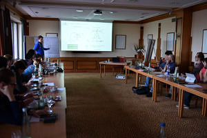
Symposium "Correlative and in situ microscopy in materials research"
1. - 4. April 2019
DPG Frühjahrstagung, Regensburg -
5th CENEM Summer School for X-ray Scattering
29. July - 1. Aug. 2019
Institute for Crystallography and Structural Physics (ICSP)
News
Annual CENEM Workshop in Bad Rodach
The annual "CENEM Workshop on Advanced Electron and X-ray Microscopy Techniques" took place in Bad Rodach during 5-7. Nov. this year. Extended presentations by our motivated colleagues provided very good overview of our on-going research. Intense discussions have stimulated sparks in new research ideas and directions.
CENEM Research on the cover of Small Methods
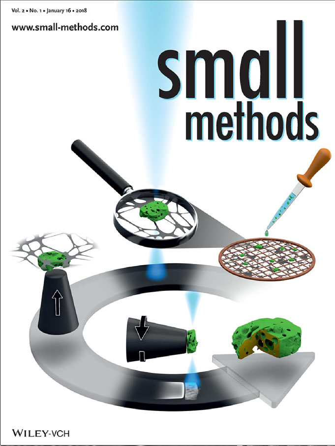
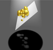
New tomography technique developed at IMN highlighted on the cover of Small Methods In the research paper Thomas Przybilla, Benjamin Apeleo Zubiri and co-workers present a versatile approach that provides a general routine for advanced 3D characterization of micro- and nanoparticles. The method which was developed at IMN and CENEM involves the transfer of individual particles onto tailored tips using a scanning electron microscope equipped with a micromanipulator. The damage- and contamination-free preparation of freestanding particles enables 360° electron tomography with unprecedented quality as demonstrated for porous zeolite microparticles and alpha-hematite nanoparticles. Find the paper here.
GRK1896 Extended
The Research Training group "in situ Microscopy" has been extended
We are glad to announce, that our integrated research training group GRK1896 has been extended for another four and a half years by the German Research Foundation (DFG)! The funding provides more than five million
euros for the training of young researchers focusing on the development of new materials using in situ microscopy and related techniques.
Read the complete press release (german only)
Mystery of butterfly research resolved
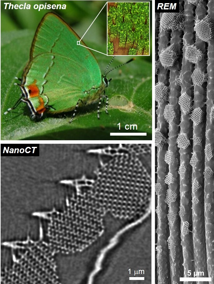
High-resolution X-ray tomography reveals how butterflies form their iridescent wing scales.
It has only been one year since the material scientists around Prof. Erdmann Spiecker from the Center for Nanoanalysis and Electron Microscopy (CENEM) at the FAU Erlangen-Nuremberg had been granted funding for one of the World's best X-ray microscopes, and they could already contribute with fascinating 3D analyses to unravel an open question in butterfly research. Their results have recently been published in the renowned scientific journal Science Advances.
Fascinating colors generated by photonic crystals
Who is not fascinated by the wonderful iridescent colors of butterfly wings? If you try to understand this phenomenon, you will find out that most often the color is not generated by pigments but rather by periodic structures made of chitin, a structure-forming polysaccharide. These so-called photonic crystals give rise to structural col-or by only reflecting specific wavelengths of the incoming solar spectrum. The re-sulting color is not random, but rather serves as camouflage or signalling. But how do millions of these photonic crystals form within the tiny scales of butterfly wings? The opinions of scientists differ in this matter.
Read more ...
Several million euros of funding for neutron and X-ray radiation research at FAU
FAU has received 2.5 million euros in funding from the Federal Ministry of Education and Research (BMBF) for five
collaborative projects on neutron and X-ray radiation research. The aim of these projects is to strengthen research
infrastructure, such as the Heinz Maier-Leibnitz Zentrum in Garching, with the FAU researchers’ expertise.
Work in this area is already a well-established part of the University’s research and involves the
faculties of Sciences, Engineering and Medicine. The DFG-funded Center for Nanoanalysis
and Electron Microscopy (CENEM) provides a platform for scattering methods to be applied in areas as diverse
as pharmaceutical excipient systems, thin films for electronics and nanostructure characterisation.
‘The new funding provided by the BMBF further strengthens this area and will allow FAU researchers
to explore new experimental methods,’ says Prof. Dr. Tobias Unruh from the Division of Crystallography and Structural Physics.
More Information on the Grant can be found
here.
CENEM Electron Tomography Expert Dr. Benjamin Winter invited to share his expertise at the Murdoch University in Perth/Australia
Dr. Benjamin Winter, an Expert in Electron Tomography at the Center for Nanoanalysis and Electron Microscopy, was invited by
the Murdoch University in Perth/Australia to give a presentation about his research. "Sharing knowledge and expertise with researchers
around the globe is an integral part of science" - Dr. Winter says -
"Therefore I am glad that I have been given the opportunity to travel to Perth to
meet many new friends and have insightful discussions about our research".
The host of the seminar, in which the talk will be given, is Senior Lecturer
Dr. Gerd Schröder-Turk, who previously
has completed his habilitation at the FAU Erlangen-Nürnberg.
More Information on the Seminar can be found
here.
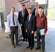
The French general consul for bavaria visits CENEM
The expertise and outstanding instrumentation available at CENEM manages to attract the attention of many people, including
diplomats like the
french general consul for bavaria. The consul was given a guided tour to the facilities of CENEM and showed a lot of interest in the
work done here.
CENEM always likes to welcome guests and demonstrate the possibility of modern-day micro- and nanocharacterization.
The ground-breaking for the new building was celebrated
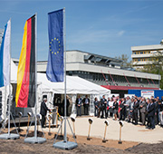
On the 6th of May the ground-breaking of the new building CENEM will based in was celebrated. The interdisciplinary center for nanostructured films as it is called will be home to over 200 scientists from different fields, strengthening the collaborative efforts in Erlangen. The building which is scheduled to be completed in 2018 has a budget of over 40 mio. € and will house several first-class laboratories for electron microscopy. Additionally the Optical Imaging Center Erlangen (OICE) will be located in the building, complementing CENEM by providing access to advanced light microscopy. CENEM is very much looking forward to its new home and new collaborations!
CENEM has been granted 3.1 Mio € for a new high-resolution x-ray microscope
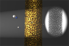
The Center for Nanoanalysis and Electron Microscopy (CENEM) has been granted funding for a new high-resolution x-ray microscope, which will enable
entirely new perspectives in materials science. Researchers will be able to analyze the 3D-structure of materials non-destructively on a sub-micrometer scale
and use the obtained information for the targeted development of novel materials.
With this technique it is for example possible to explore the tiny pore structure of nano-foams and optimize them for catalytic applications.
More info will follow soon!
The new cryo transfer holder for electron tomography has arrived
CENEM is proud to announce that the first TEM holder of its cryo-TEM subdivision has arrived. The Fischione cryo transfer holder enables the transfer of frozen samples into the TEM while holding a constant temperature of -196° C, enabling the analysis of biological and especially fragile samples. In addition to conventional analysis the holder also enables tomographic analysis having an extended tilt range of +-78°. With the sample preparation having already been established at CENEM the cryo-TEM division can begin its exciting work.
Logging activities in preparation for the construction of the new building for CENEM
The cluster of excellence is planning the construction of a new building which will house new laboratories for CENEM as well as the
Optical Imaging Center Erlangen (OICE),
which is a complementary center for characterization using advanced light microscopy.
The intial logging activities are already
completed making way for the laying of the foundation in early 2016. The building will cost about 40 million Euros and is scheduled
to be finished in 2018. Besides the two strong centers for characterization there will be a focus on nanostructured films represented
by several groups from the FAU. CENEM is very pleased that several high-end laboratories for nanocharacterization are planned, ensuring
future growth of the core facility. Stay tuned for future updates on the progress of the construction!
FAU researchers answer key question on the microstructure of butterfly wings using electron tomography at the Center for Nanoanalysis and Electron Microscopy
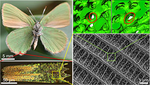
FAU researchers have succeeded in pinpointing
the three-dimensional structure of microscopic
photonic crystals in butterfly wings.
These findings will help researchers to
understand the formation of periodic
structures which affect the colouration
of many butterflies. In the future, these
findings could be pivotal in designing new optical materials.
The research findings which originated from a close
collaboration within the Cluster of Excellence
‘Engineering of Advanced Materials’ between the
FAU-based Institute of Micro and Nanostructure Research
and the Chair for Theoretical Physics 1 (Prof. Dr. Klaus Mecke),
as well as the School of Engineering and Information Technology at
Murdoch University Australia (Prof. Dr. Gerd Schröder-Turk) have
been published in the prestigious interdisciplinary journal
Proceedings of the National Academy of Sciences of the United States of America (PNAS).
Read the full story on the news page of the FAU.
Visitor numbers at the Long Night of Science exceed all expectations
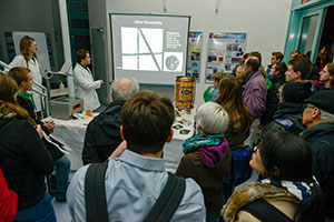
The Long Night of Science was a huge success for CENEM. At the Electron Microscopy facilities alone we welcomed more
than 400 people throughout the night. Visitors had the chance to see the new Titan Themis3 and learn about research
at the Institute for Micro- and Nanostructure Research in a vivid presentation. Several demonstrators were on display, including a
mini gas turbine and a self-cooling beer keg illustrating the impact of research on everyday life.
Visitors were also invited to try warm wine and beer from the self-cooling kegs, kindly provided by Tucher.
In the end everyone involved in the event can be proud to have helped convey the importance of science to the general public.
We are looking forward to next time!
Researchers at the FAU develop a novel spin-crossover complex - with help from the research training group "in-situ microscopy"
Metal-organic spin-crossover complexes exhibit the unique possibility to
switch between two distinct states with high or low total spin. They thus
represent promising materials for a new generation of functional devices
which take advantage of this phenomenon at the molecular scale. Scientists
at the FAU (Friedrich-Alexander Universität Erlangen-Nürnberg) now published
investigations on a specifically developed spin-crossover complex in the journal
“Angewandte Chemie” (DOI:10.1002/anie.201504192). This complex is
able to undergo the spin transition from a paramagnetic (magnetic) state in a
diamagnetic (non-magnetic) state and reversely triggered by light
irradiation at room temperature and in the solid state.
Read the full press note here.
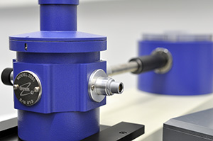
Installation of VAXSTER
On March 18th 2015 the upgrade of our unique SAXS/GISAXS instrument VAXSTER (Versatile Advanced X-ray Scattering instrumenT ERlangen) started. This includes the full rebuild of the primary instrument part with the 3/4 slit collimation system focusing X-ray mirror and the X-ray source. VAXSTER will become an extremely versatile combined SAXS/GISAXS/GIXD laboratory instrument with outstanding performance. It will be equipped with a MetalJet D2 70 kV X-ray source from EXCILLUM, Sweden. It allows for horizontal and vertical grazing incidence measurements within a fully evacuated setup or with a sample positioned at ambient conditions. The beam will be shaped by a 150 mm Montel optics (INCOATEC, Geesthacht) and a set of up to 4 double slit systems with the last two slit systems equipped with low scattering blades (JJXray/SAXSLAB). Here you will find some photographs of the ongoing installation of VAXSTER.
Installation of the new Titan Themis³ 300 TEM
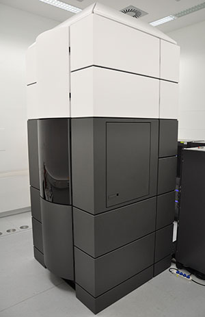
In October 2014 the installation of the new transmission electron microscope (TEM) FEI Titan Themis³ 300 was finished. Now the staff of CENEM is able to perform high quality research using this high-end TEM providing high-resolution imaging as well as high performance analytical investigation capabilities. This double-aberration-corrected TEM corrects both the image and the probe forming system enabling high-resolution imaging in TEM as well as scanning TEM (STEM) mode resulting in resolution limits below 1 Å for both modes for all high tensions between 60 kV and 300 kV. The high brightness electron gun (X-FEG) equipped with a monochromator to improve the energy resolution in combination with a high-sensitivity SDD X-ray spectrometer (Super-X) and a high-resolution post-column energy filter (GIF Quantum) creates a high performance analytical instrument perfectly suited for the nanoanalytical characterization of a variety of materials and devices. The FEI Titan Themis³ 300 was installed instead of a FEI Titan³ 80 – 300 TEM to improve the electron microscopical performance and investigation capabilities of CENEM.
New chair “Institute of Micro- and Nanostructure Research”
The group Electron Microscopy, that builds one part of CENEM, was assigned to the Institute for Biomaterials at the Department of Materials Science and Engineering of the University of Erlangen-Nürnberg. This working group evolved significantly in the last years in terms of staff members and scientific output. Therefore a new chair was established for this promising Electron Microscopy group in autumn 2014. The new chair is called Institute of Micro- and Nanostructure Research and is assigned to the Department of Materials Science and Engineering of the University of Erlangen-Nürnberg. The inauguration of the new chair will be celebrated together with the inauguration of the new FEI Titan Themis³ 300 TEM on the 30th of April 2015.
EMS Outstanding Paper Award
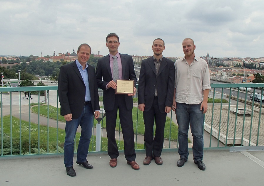
The European Microscopy Society (EMS) awards each year Outstanding Paper Awards in the three categories Instrumentation and Technique Development, Materials Sciences as well as Life Sciences. In 2013 the publication "Dislocations in bilayer graphene" wins the EMS Outstanding Paper Award in Materials Sciences. We congratulate the authors Benjamin Butz, Christian Dolle, Florian Niekiel, Konstantin Weber, Daniel Waldmann, Heiko B. Weber, Bernd Meyer and Erdmann Spiecker, who published this paper in the journal Nature. The award took place within the 18th International Microscopy Congress 2014 in Prague.