

Current Events:
-
2nd Erlangen School on Atom Probe Tomography
4. - 8. March 2019
Institute I, Materials Science and Engineering -
GRK 1896 Springschool
18. - 20. March 2019
Pommelsbrunn -
Symposium "Correlative and in situ microscopy in materials research"
1. - 4. April 2019
DPG Frühjahrstagung, Regensburg -
5th CENEM Summer School for X-ray Scattering
29. July - 1. Aug. 2019
Institute for Crystallography and Structural Physics (ICSP)
Scattering Methods Facility
The team of the nanomaterials characterization work group
uses advanced
scattering methods to characterize nano-structured materials. In this
context we study structure formation in complex systems, the
stabilization and degradation mechanisms in colloidal solutions and thin
films mainly by small angle scattering techniques (SAXS, SANS, GISAXS)
but also by WAXS and GIXD. The main scientific laboratory instruments of
our research group are the state-of-the-art SAXS/GISAXS machine VAXSTER
with unique features and outstanding performance and the high throughput
SAXSpace instrument.
Small angle X-ray scattering
Versatile Advanced X-ray Scattering instrumenT ERlangen (VAXSTER)
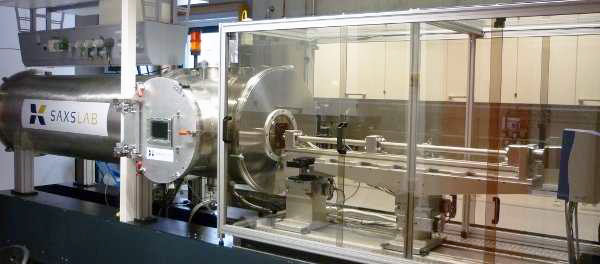
- Source: Xenocs Genix 3D beam delivery system
- ultra low divergence
- collimating
- beam size (FWHM) of 1.1x1.5 mm²
- divergence (FWHM): 0.4 mrad
- Collimation: beam shaping: source multilayer optics, collimation through 3 sets of 4-bladed slits
-
Detector:
- Pilatus 300K detector (500 Hz):
- three modules à 83.8x33.5 mm², 487x619 pixel à 172x172 μm²
- readout time of 3 ms
- energy range of 4.5-36 keV at a resolution of 500 eV
- Sample positions: under vacuum (~10-5 bar) or ambient conditions (large space for mounting different sample stages)
-
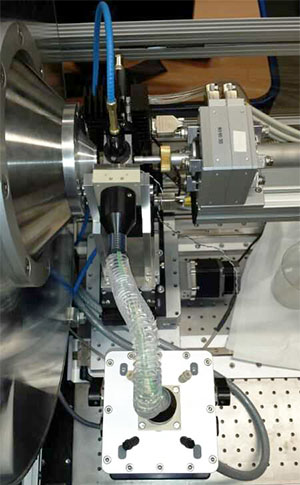
Sample stage:
mounting of various sample stages inside detector tube possible (i.e. Measurement in vacuum), as well as externally (measurement in air/controlled vapor environment), yz-theta goniometer for transmission as well as grazing incidence applications Many different sample holders for mounting on yz-sample stage including:- GISAXS sample holder
- temperature-controlled Cu-holder with 5 sample-positions
- Stopped-flow cell
- capillary holder
- Q-range: Total Q-range: Q = 0.003-4Å-1 low count rates for small Q-limit, only achievable under ambient conditions; high Q-limit only achievable in vacuum individual configurations in between; e.g.: WAXS (0.05-2.5 Å-1), MAXS (0.012-0.67 Å-1), SAXS (0.006-0.26 Å-1), Extreme-SAXS (0.003-0.21 Å-1)
- spec
SAXSpace (Anton Paar)
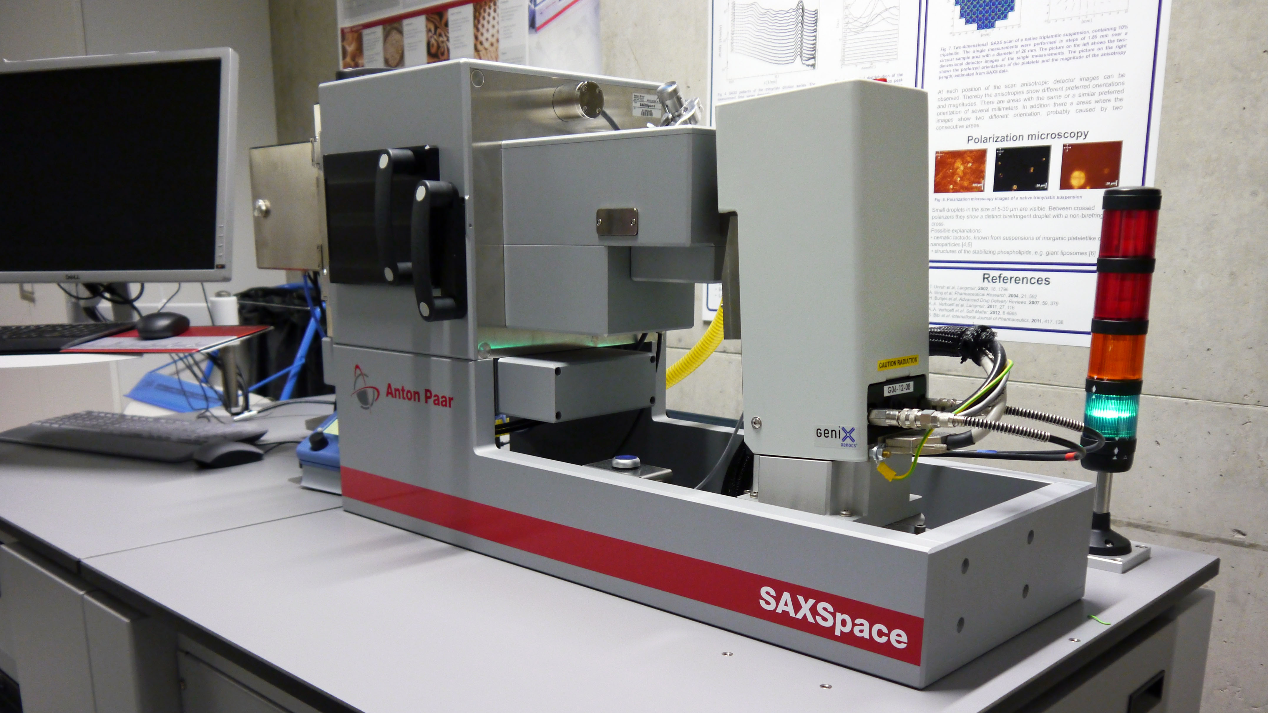
- small angle (SAXS → approx. 0.04° to 10°) and wide angle (WAXS, up to 74°) X-ray scattering
- X-ray source: 50 W Cu Kα microfocus tube 2D Diode-array (CMOS) detector (Pilatus 100k)
- focusing multilayer mirror to monochromatize and enhance X-ray intensity
- scatterless Kratky-based collimation system for line and point collimation
- Sample holders for liquids, pastes and solids
- temperature range of -30 °C to +150 °C. (peltier heating/cooling)
- SAXSdrive, SAXStreat and SAXSquant software
Stopped-Flow SFM-2000
- two 10 ml syringes
- minimal injection volume per syringe: 28 μl
- flow rate from 0.062 – 10 ml/s per syringe
- minimum total flow rate for efficient mixing: 1 ml/s
- ratio range from 1:1 to 1:40
- duration of flow: 1 ms to 60 000 ms per phase
- minimal dead time 0.7 ms
- Berger ball mixer
- Bio-Kine 32 V4.66 for syringe control
X-ray diffraction
Philips/PANalytical X’Pert Pro-MPD
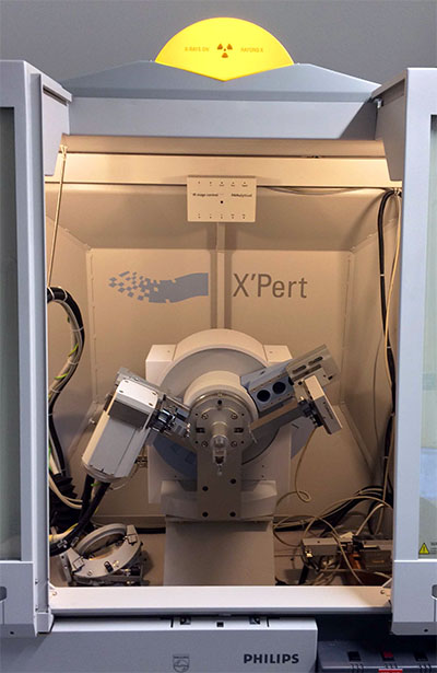
- Powder Diffractometer
- Bragg-Brentano-Geometry (2Θ ca. 10° - 100°) or
- Gracing-Incidence Powder Diffraction (GID, incidence angle>≈0.2°)
- Anode Cu (Kα)
- Running Parameter 40kV, 35mA
- Filtering of Kβ
- Soller Collimation
- Semiconducting stripe detector (X'Celerator) or
- Gas ionisation counter (Miniprop)
- Variable step size
- Automatable
- optional:
- Reflectometry stage
- Eulerian Cradle
- Capillary optics
Huber Guinier Diffraktometer
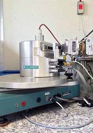
- Powder Diffractometer
- Transmission-Geometry (2Θ fixed 10° - 100°)
- Anode Cu
- Monochromator: Kα1
- High angular resolution
- Running Parameter 40kV, 35mA
- Illumination time: ca. 10min - several hours
- Image plate online read-out
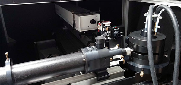
Rotating Anode Setup for Powder Diffraction:
- Rotating copper anode
- Crossed goebel mirror setup for monochromatic Cu Kα radiation
- Voltage and current up to 44 kV and 75 mA, respectively
- Measurement in transmission Debye-Scherrer geometry
- 2D ccd detector allows measurement of full diffraction rings
- 2θ range of 0° to about 50°
- In-Situ chamber for variation of temperature ramps up to 550° C
- Eurotherm controller
- Possibility for different atmospheres (vaccum, inert gases)
- 6 axis goniometer for adjustment of sample position
Thermoanalysis
Micro DSC III Analyzer (Setaram)
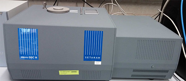
- High sensitivity DSC and calorimetry
- Temperature range: -20 to 120 °C
- Scanning rate: 0.001 to 1.2 °C/min
- Vessel volume: 850 µl
- Resolution: 0.3 µW
- Cooling rate from 120°C to room temperature: 1.5 h
- Scanning or isothermal mode
- Thermal Analysis Software: Calisto
Perkin Elmer DSC 8500
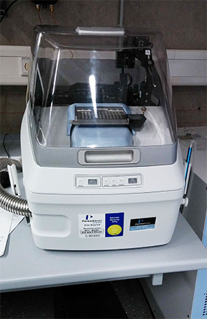
- Hyper-enabled Double-Furnace Differential Scanning Calorimeter
- Cooling system: Intracooler 3 (Perkin Elmer), three stage, closed- loop circulationg heat exchanger
- Temperature range: -100 to 750 °C (by nitrogen usage)
- In-situ ballistic cooling to 2100 °C/min
- Scanning rate: 0.1 to 750 °C/min
- Sample pans: aluminium (50 µl)
- Analysis Software: Pyris
Light scattering and spectroscopy
BI-200SM Goniometer (Brookhaven Instruments Corporation)
- motor driven angle selection (0.01° steps)
- narrow-band optical filters for laser wavelegths of 633, 514.5, 488 nm
- 100, 200, 400 μ pinholes for selecting coherence areas (QELS measurements)
- 1, 2, 3mm apertures for adjusting intensity (classical measurements)
- Selected photomultiplier tube (PMT) as Detector
- circulating index matching liquid with dust filter
- Temperature controle approximately+5°C to 80°C
- needed sample Volume: 1.5mL (strongly diluted)
- Diode Laser 637 nm (BIC), 30mW
- Lexel95-2 (Lexel), Argon Laser, 476.5 nm – 514.5 nm, 4 W, 1.33 mm beamdiameter, 0.6 mrad beam divergence
UV-Vis Spectrometer TIDAS S 500K
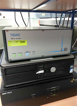
- - wavelength range 187.79 – 1016.53 nm
- spectral resolution < 2.5 nm
- wavelength accuracy < 1.0 nm
- wavelength reproducibility 0.1 nm
- baseline drift at 250 nm: 5 μAU/h
- signal-to-noise ratio < 20 μAU
- integration-time range 0.7 – 10 000.0 ms
- integrated D2- & halogen light source
- detector MCS - Aspen 780 kHz
- photodiode array: 1024 pixels
- Bio-Kine 32 V4.66 for data acquisition and analysis